Shape Analysis Group
Conduction in the Heart Wall
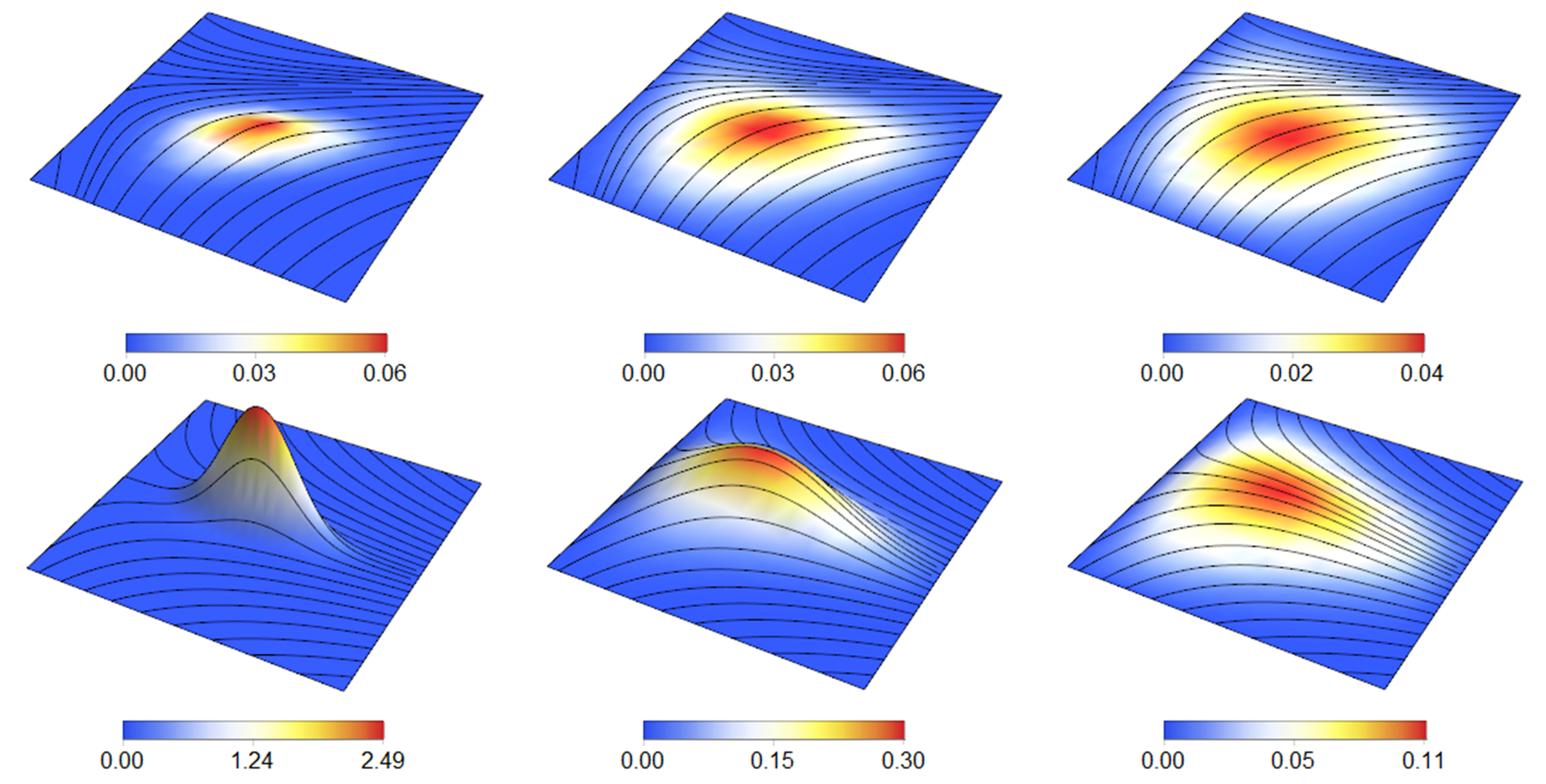
The mammalian heart must function as an efficient pump while simultaneously conducting electrical signals to drive the contraction process. In the ventricles, electrical activation begins at the insertion points of the Purkinje network in the endocardium. How does the diffusion component of the subsequent excitation wave propagate from the endocardium in a healthy heart wall without creating directional biases? We show that this is a consequence of the particular geometric organization of myocytes in the heart wall. Using a generalized helicoid to model fiber orientation, we treat the myocardium as a curved space via Riemannian geometry, and then use stochastic calculus to model local signal diffusion. Our analysis shows that the helicoidal arrangement of myocytes minimizes the directional biases that could lead to aberrant propagation, thereby explaining how electrophysiological principles are consistent with local measurements of cardiac fiber geometry. We discuss our results in the context of the need to balance electrical and mechanical requirements for heart function.
Related Publications
Tristan Aumentado-Armstrong, Amir Kadivar, Peter Savadjiev, Steven W. Zucker and Kaleem SiddiqiConduction in the Heart Wall: Helicoidal Fibers Minimize Diffusion Bias.
Scientific Reports, 8(7165), 2018.
[online]